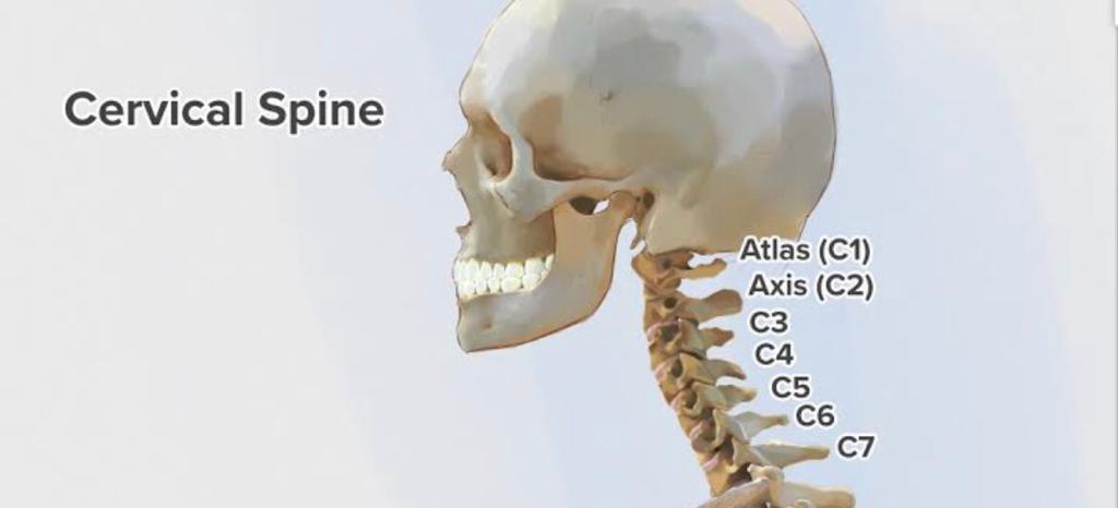
This is the uppermost part of the spine and is composed of 7 vertebraes. This is devided in to two groups
Important note:- Three joint segment
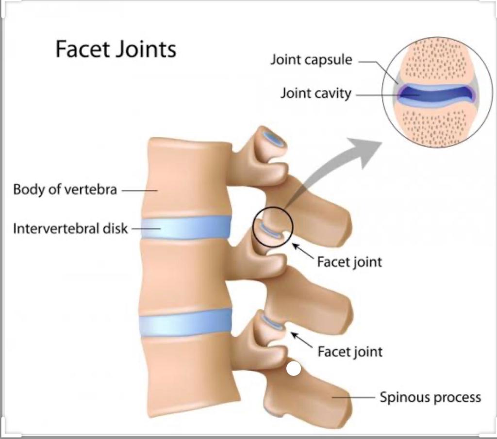
At any one level,the motion segment is composed of three distinct points
The two facet joints and one intervertebral joint.They are both interdependent on each other.
Let me explain
So any abnormality in facets affects the disk and any variation in the disk height affects the facets So, in spine it is always better to think more in terms of segment dysfunction rather then joint dysfunction.
Risk factors are following
1) poor postures while using laptops, mobiles, studying etc
2) sleeping in bad positions
3) lack of proper diet and exercises
Can cause “ CERVICAL INSTABILITY “
Normally cervical spine stays in slight lordosis, which provides passive stability from facets and ligaments Too much bending causes
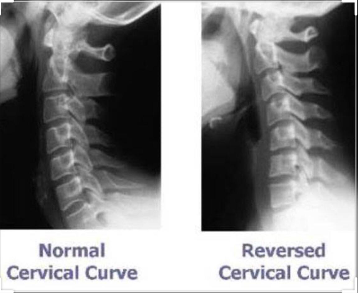
Reversal of lordotic curve
Passive stability is lost and the segmental muscles must go in to constant contraction which causes muscle spasm and inflammation. Also too much stress on intervertebral joint and decrease in disc height
a) Disc disease (Acute or chronic)
b) Spondylosis (degenerative joint disease
Disc disease(Acute or chronic)
If the annulus of the disk is weakened, it can result in eithe anterior or posterior disk bulge Anterior disk bulge presses on the anterior ligaments and muscles and a patient may feel difficulties in swallowing or have a sensation of sore throat. Posterior disc bulge can press the posterior muscles and ligaments along with spinal cord or spinal nerves.
Symptoms
Neck pain, usually noticed early in the morning. It has been described as acute dislocation of the facet joint or entrapment of the synovial villi. The pin begins with clearly delineated sharp neck pain and progresses during the day to generalised muscle spasm and inability to rotate the neck in one direction.Typical presentation is neck rotated and bent to one side and acute distressful pain.Usually traction in extension given at this time relieves the painful symptoms immediately.
Degenerative joint disease(spondylosis)
This begins as capsular restriction of the facet joints and with out any bony changes on Xrays.Gradually progresses over months or years to
The flattening
|
Lipping
|
Spurring of the vertebral bodies
This whole process is considered as normal aging process (in old people) but it should be considered abnormal in younger people.Read Above(Cervical instability)
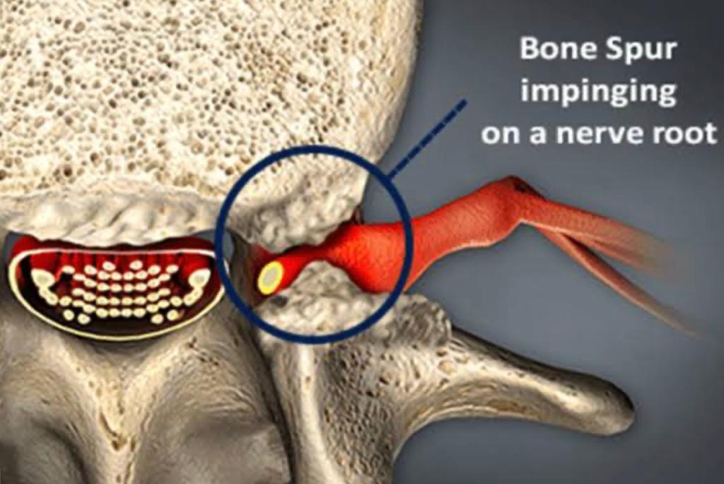
A) Bony stenosis of the intervertebral foramen and result in neck pain,shoulder pain, radiating pain in the arms/forearm till fingers,paraesthesia/numbness and muscle weakness.
Patient presentation
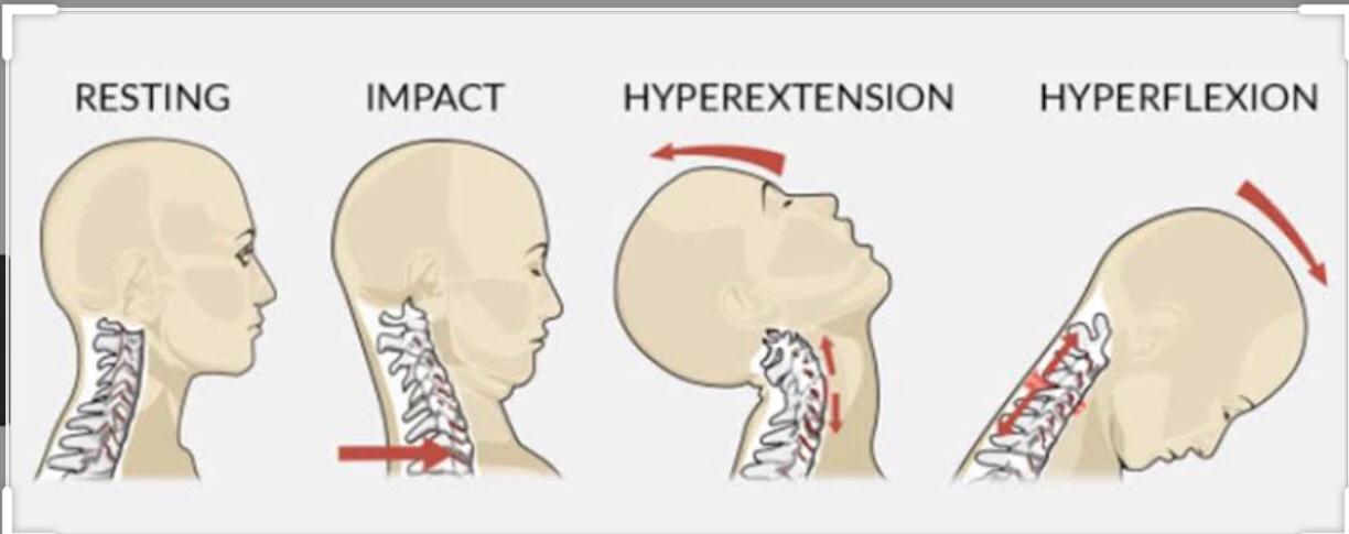
The mechanism of acceleration injury is acceleration of the head and neck relative to the body.It results from the collision of two automobiles but also can result contact sport such as football and high-velocity sport such as skiing.
Mechanism of injury is a stationary automobile struck from behind.At the moment of impact,the trunk of the body, which is supported by the car belt, moves rapidly forward.The moment of inertia of the head creates a relative backward accelerationof head and neck.
Acceleration injury should be considered in following three phases
1) Acute
2) Subacute
3) Chronic
Acute phase
This starts at the moment of the accident and lasts as long as 2 to 3 weeks.There is generally little pain and fairly free ROM immediately after the accident with painful stiffness gradually developing after 24 to 48 hours.
There is a possibility of
A) fracture (fracture injury to the nerve roots)
B) Contusion to the spinal cord
C) Head injury
D) Cervical sprains and strains
Subacute phase
This lasts 2 to 10 weeks.The muscle pain originally experienced is gone and is replaced by deep,aching pain that may be referred to head,interscapular area,or the upper extremity. An X ray taken at this time may show flattening due to muscle spasm.
Chronic phase
This begins when the acute healing process is over and the larger muscle groups have completely healed but they may be shortened and fibrotic. Because of ongoing spasm of the longus colli (Anterior neck muscle) The cervical spine can go in to Read Above (Cervical instability)
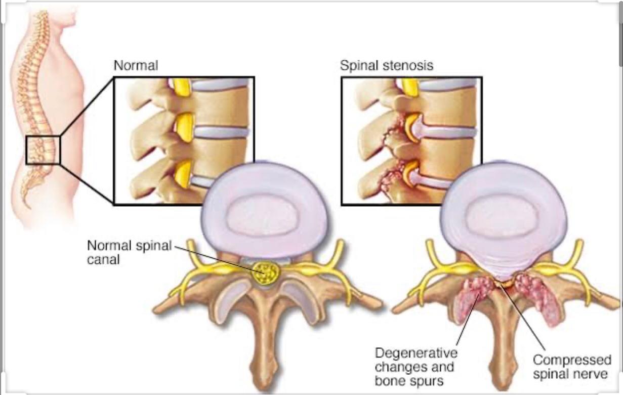
This includes narrowing of the spinal canal,nerve root canals and intervertebral foramina, all of which causes nerve root entrapment.
Presentation :-
Back Pain Transient Motor Deficits
Tingling and intermittent pain in one or both legs which is worsened by standing or walking( Neurogenic claudication) and some what relieved by sitting
(Difference between vascular and ***neurogenic claudication is that the relief experienced at rest in vascular is rapid as compared to neurogenic which may take few hours)
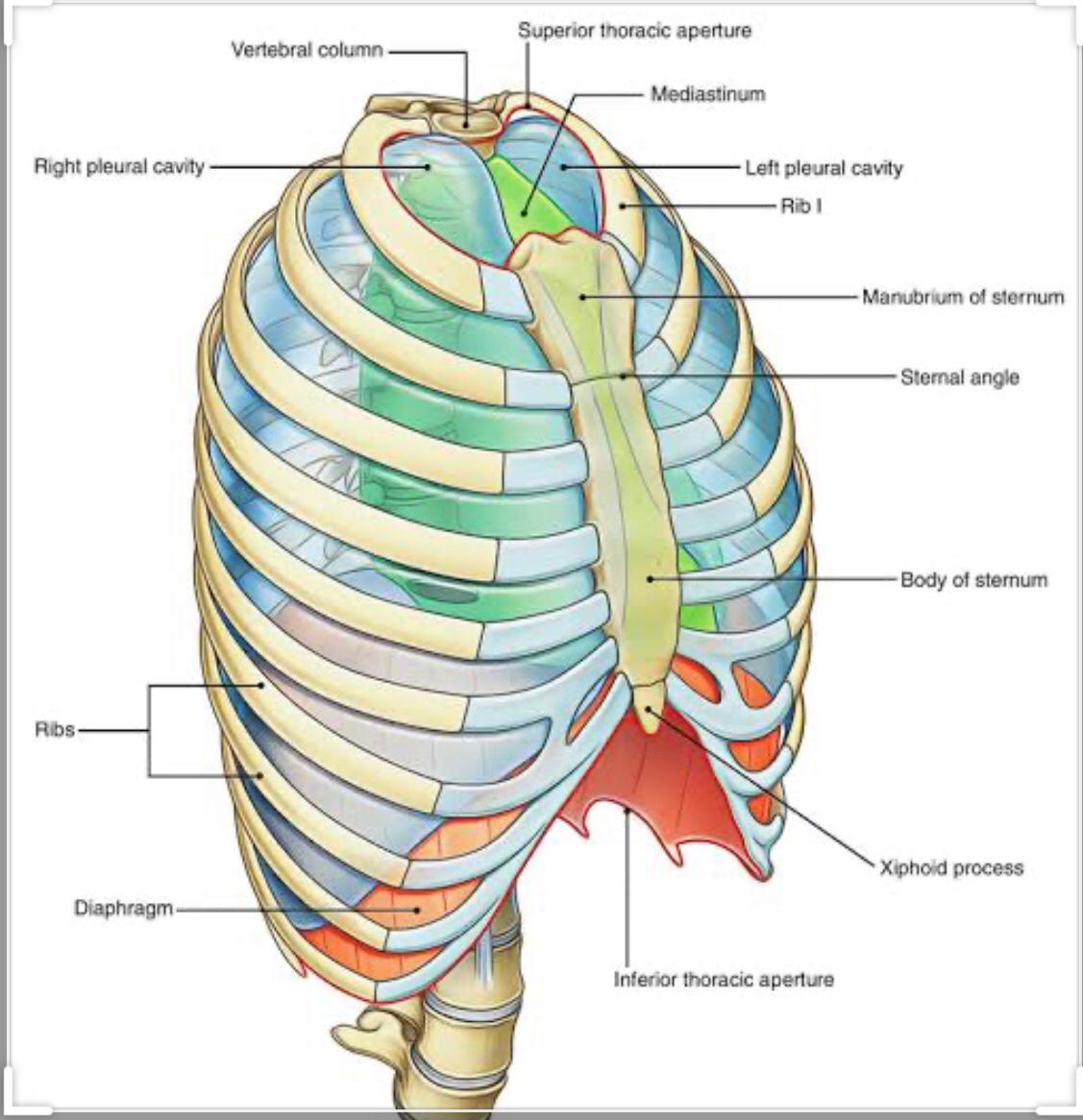
It consists of relatively rigid rib cage formed by the 12 ribs, 12 thoracic vertebraes and sternum. Important functions
1) stable base for muscle to control cranio cervical region
2) protection of inner thoracic organs
3) mechanical bellows for breathing
A) Thoracic disc herniations Of the total number of disk problems, thoracic presentation is 1 to 6 patients per 1000.The problem appears predominantly in males and is common in the 5th decade of life. T11 -T12 are most commonly involved.It can result in PARAPLEGIA
B) Minor intervertebral dearangements (MID) It is defined as “isolated pain” in one intervertebral segment of mild character due to mechanical cause Patient presentation.It always involves one or two facet joints in the mobile segment,thus initiating novice price activity in the posterior primary dermotome and myotome.
Patient presentation
a) local tenderness and muscle spasm
b) reffered pain Reff
Read Above(Three joint segment)
C) Thoracic pain of lower cervical origin
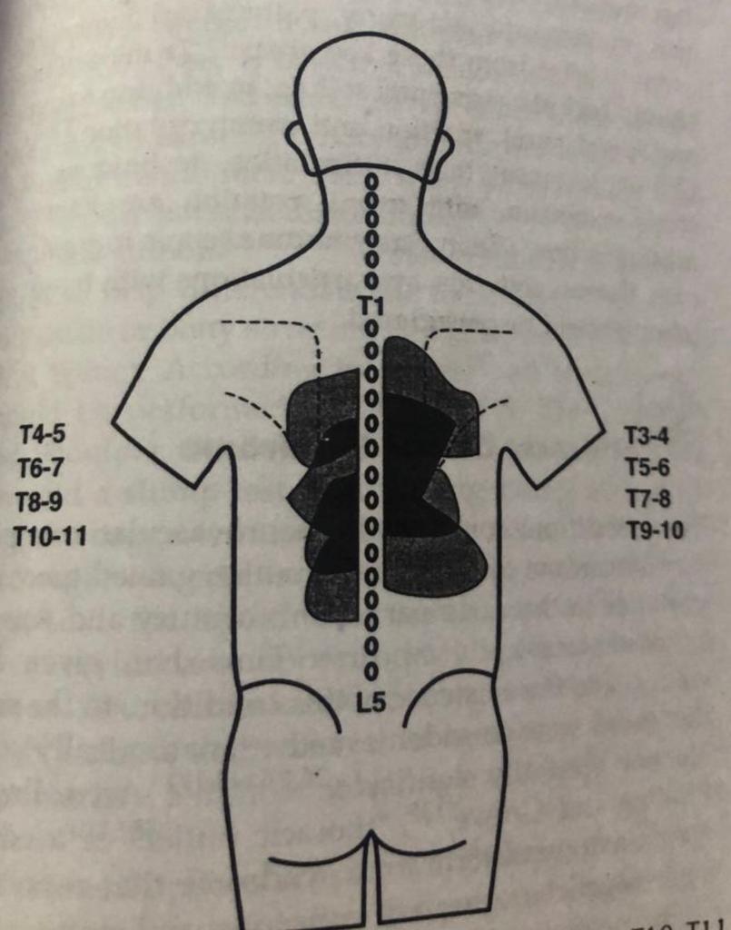
Any MID in lower cervical region C5C6 ,C6C7 and C7 T1 can cause reffered pain in the interscapular point near T5 T6
D) Thoracic pain of thoracic origin
E) Thoracic hypomobility syndrome
This results from fibrosis (capsular) of the facets resulting in the decreased height of the disc due to lack of nutrition
According to McKenzie :-
Extension dysfunction develops in the patients with both sheurmann’s disease and osteoporosis NOTE ON OSTEOPOROSIS:-
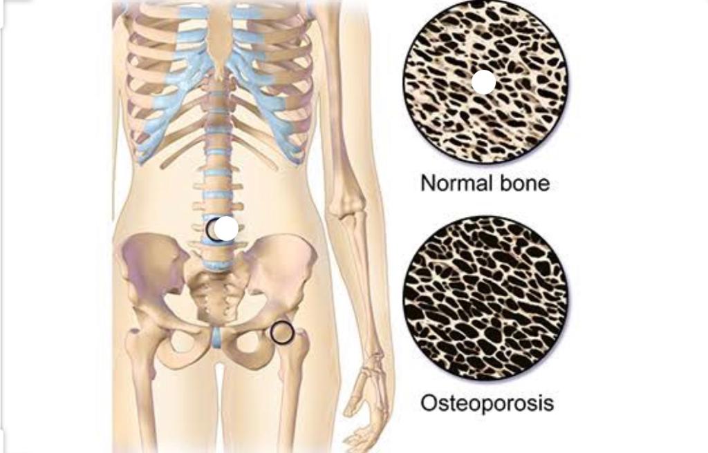
It results from lower then normal maximum bone mass and greater than normal bone loss.This results in the kyphosis deformity in the dorsal spine.Commonly occurs in women with menopause due to decrease levels of estrogen.
It also occurs in number of diseases and treatments
Myoclinic research in post menopausal spinal osteoporosis has demonstrated that extension exercises performed regularly significantly reduces osteoporosis and number of compression fractures
F) Thoracic outlet syndrome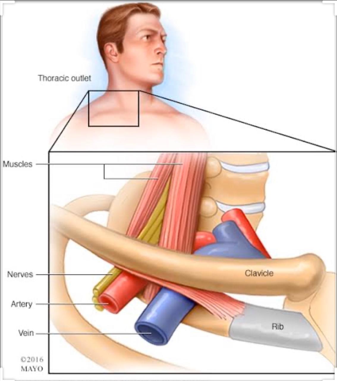
Neurovascular compression syndrome of the thoracic outlet It occurs due to combined anomalies of the soft tissues and the bony boundaries of the outlet. Example
Bony boundaries:- Cervical ribSoft tissue :- anterior scalene syndrome,pectoralis minor syndrome
G) Rib conditions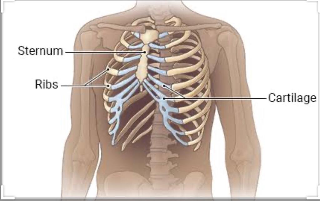
Some painful conditions are associated with small derangements of the Costco-vertebral or Costco-transverse articulations
Let me explain:-Twelve pairs of ribs enclose thoracic cavity forming a protective cage for cardio-pulmonary organs.They connect anteriorly to the sternum and posterioly form 2 joints
a) Costo vertebralAny strains or sprains in the ribs or joints cause painful condition
Hypermobility or hypomobility of the ribs or joints also cause painful condition
H) Scoliosis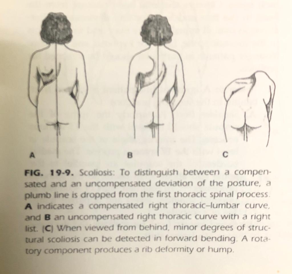
This is a medical condition in which a person’s spine has a sideways curve.
S shaped or Cshaped Over three dimensions Classification
1) Structural in which spine is fixedA structural scoliosis doesn’t straighten during forward bending or side bending in to the convexity of the spine. Because side bending is always accompanied by rotation, there will be a lumbar bulge or rib hump(gibbus) with forward bending if a structural scoliosis is present.
A functional scoliosis can be caused by muscle imbalance, poor posture or a leg length discrepancy.This kind of scoliosis generally straightens on forward bending and side bending towards the convexity, except in the presence of muscle spasm or guarding.
A note on list Facet joint impingement or nerve entrapment in the intervertebral foramen may also cause pelvic deviation to one side and lumbosacral deviation to same or opposite side depending upon the site (medial to nerve root or lateral)
This marks as the second most common reason to visit Opd after common cold.
According to Dagi and beary about 80% of all back cases can be attributed to soft tissue conditions ie. strains, sprains , postural abnormalities, poor muscle tone or neuromuscular diseases and 10% are because of intervertebral diseases with or without radiculopathy and remaining10% due to serious causes eg Timor’s.
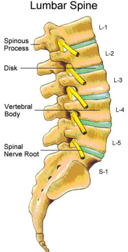
Read Above Reff 3 joint segment* Facet (zygapophyseal joint)
In lumbar region,degeneration of the facet joint is a rule rather than an exception in virtually everyone living beyond third decade or so..The fact is attributed in part to the likelihood that human spine has not completely adopted to the upright,weight bearing position and to the fact that human life expectancy has been drastically extended.
Degeneration in the lower back initiates with
1st scenarioPerson complains of “stiff back- tissue gradually adapts by means of fibrosis and bony hyper trophy - radiologically not very drastic changes are visible
2nd scenarioPerson complains of pain due to acceleration of degenerative process of the spinal joints due to
In these cases tissues fail to adopt and pain initiates relatively early
Vertebrae
Intervertebral discs
This marks as the second most common reason to visit Opd after common cold.
According to Dagi and beary about 80% of all back cases can be attributed to soft tissue conditions ie. strains ,sprains , postural abnormalities, poor muscle tone or neuromuscular diseases and 10% are because of intervertebral diseases with or without radiculopathy and remaining10% due to serious causes eg Tumor.
Reff 3 joint segment* Facet (zygapophyseal joint)
In lumbar region,degeneration of the facet joint is a rule rather than an exception in virtually everyone living beyond third decade or so..The fact is attributed in part to the likelihood that human spine has not completely adopted to the upright,weight bearing position and to the fact that human life expectancy has been drastically extended.
Degeneration in the lower back initiates with
1st scenarioPerson complains of “stiff back- tissue gradually adapts by means of fibrosis and bony hyper trophy - radiologically not very drastic changes are visible
2nd scenarioPerson complains of pain due to acceleration of degenerative process of the spinal joints due to
In these cases tissues fail to adopt and pain initiates relatively early
Degenerative conditions of Lower back
Resulting in the abnormal function of the posterior element and disk DAMAGE IS REVERSIBLE
2) Intermediate “Instability”This is a stage of laxity and capsule and annulus becomes hyper mobile Restabilization of the structure results in the formation of osteophyte and traction spurs.
3) Final stabilisation phaseFibrosis of the posterior joints and capsule ,the loss of disk material Osteophyte formation around the 3 joint segment increases the load bearing surface and decrease motion resulting in a stiff painful motion segment.
Resulting inEnquire Now
